From the Bench: Creative Approaches in Cancer Research
Improved patient outcomes come from new treatments. New treatments stem from successful clinical trials. Clinical trials build on discoveries made in the lab.
And laboratory discoveries? Those rely on a vast array of assays and specialized technologies, combined with the ingenuity of bench scientists who devise, master, and apply these tools in creative ways to investigate scientific questions.
Advances in the approaches used by researchers, therefore, have far-reaching impact—enabling cutting-edge studies and novel insights into cancer that could ultimately benefit patients.
To help readers stay abreast of new tools and strategies, Cancer Research Catalyst is launching a new quarterly series, “From the Bench,” to highlight some of the creative approaches researchers are employing in the lab to understand cancer and to improve its detection and treatment.
Our first installment features cancer-smelling insects, new strategies for delivering proteins and modulating hypoxia in cells, and a self-charging battery that can fight cancer.
Coaxing tumor cells to collaborate with the immune system
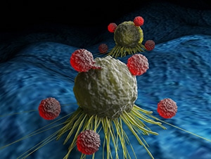
What if cancer cells could be reprogrammed into antigen-presenting cells (APCs) that can stimulate anticancer T-cell immunity? In a study published in the AACR journal Cancer Discovery, a team of researchers showed that this approach may be feasible and could lead to a new strategy for therapeutic cancer vaccination.

In this study, researchers induced expression of master myeloid transcription factors in murine leukemia and solid tumor cell lines to trigger lineage reprogramming of these cells into APCs. The tumor-reprogrammed APCs, named TR-APCs, expressed myeloid markers and exhibited immunostimulatory functions.
When B-cell leukemia TR-APCs were transplanted into mice, turning on the expression of the myeloid transcription factors during tumor development resulted in the eradication of leukemic cells and prolonged survival compared with mice transplanted with non-reprogrammed leukemic cells. This antitumor effect was associated with expansion of activated and memory T-cell populations and reduction of immunosuppressive regulatory T cells. Tumor eradication and improved survival was also observed with TR-APC models of solid tumors.
To explore the translational potential of the TR-APC approach, the researchers reprogrammed human B-cell acute lymphoblastic leukemia blasts by stimulating them with myeloid cytokines in vitro. Importantly, the reprogrammed leukemic cells were capable of inducing the activation of autologous T cells, indicating that patient-derived TR-APCs may be used as a novel immunotherapy approach to stimulate leukemia-specific T-cell immunity.
Multi-use microcapsules fine-tune oxygen concentrations in cell culture
Cell culture incubators provide a single concentration of oxygen to all cells, and it is difficult to maintain steady and physiologically relevant levels of hypoxia in vitro when modeling hypoxia-dependent processes, such as angiogenesis and tumor cell interactions.
In one study published in the Proceedings of the National Academy of Sciences, researchers developed gelatin-based microcapsules that function as oxygen-consuming factories and can be suspended in liquid media or embedded in a variety of cell-supporting hydrogels. The microcapsules can be loaded with various concentrations of the enzymes glucose oxidase and catalase, which, combined, consume oxygen and glucose and release reactive oxygen species (ROS). The microcapsule pores are small enough to allow substrates and products in and out while trapping the enzyme, protecting it from proteases and, if the capsules are embedded in a hydrogel, allowing spatial control of oxygen concentration.
Microcapsules loaded with varying enzyme concentrations exhibited a dose-dependent effect on the growth and viability of two-dimensional melanoma cell cultures and breast cancer microspheres. Researchers also demonstrated the ability to mount a microsphere-loaded hydrogel in one area of a culture, creating an oxygen gradient ranging from hypoxic conditions near the hydrogel to normoxic conditions far from it. The authors of the study suggested that this technology may be especially useful for the coculture of cells that require different optimal oxygen concentrations, such as in “body-on-a-chip” devices.
A self-charging saltwater battery for antitumor therapy

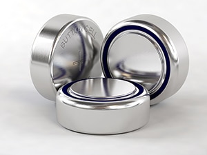
In a recent study in Science Advances, scientists investigated whether a small, implantable battery can help fight cancer. The battery charges by consuming oxygen, kickstarting a chemical reaction that releases ROS, which can damage tumor tissue. The depletion of oxygen also creates a severely hypoxic environment with an average hemoglobin-bound soluble oxygen concentration of 1.9%, compared with 14.1% without the battery.
Researchers implanted the batteries near tumors in mice and treated the mice with tirapazamine, a hypoxia-activated prodrug. The battery plus tirapazamine eliminated tumor xenografts by day 14 in 4 of 5 mice, with an average tumor volume reduction of 90%. No significant changes in body weight or major organ function and histology were observed during treatment. The researchers suggested that their battery produces a more continuous state of hypoxia than oxygen-depleting drugs, photodynamic therapy, and sonodynamic therapy, with fewer side effects.
Repurposing a bacterial “syringe” for targeted drug or protein delivery
Selective delivery of drugs, proteins, or other molecules to cancer cells is a key component of both cancer research and cancer treatment. In the lab, it helps researchers probe basic cellular processes to understand cancer; in the clinic, it helps target cancer cells while minimizing the effect on healthy tissue.
An article published earlier this year in Nature describes a new strategy for delivering drugs or proteins to cells. In this study, researchers modified a naturally occurring protein complex that is normally used by bacteria to deliver toxins or host-modulating proteins directly into target cells.
The complex, known as a contractile injection system, functions similarly to a syringe and relies on an inner tube that holds the payload and a sharp spike protein that is pushed through the membrane of the target cell. By engineering the contractile injection system of the bacterium Photorhabdus asymbiotica to recognize different cellular receptors, the researchers were able to guide delivery of various payloads to specific cell types, both in culture and in live mice.
The proof-of-principle experiments demonstrated that the modified system could be utilized to selectively deliver cytotoxic drugs to cancer cells and to direct gene editing proteins such as Cas9 to specific cells, suggesting that this novel delivery system has potential therapeutic and research applications.
A new method to engineer T cells for immunotherapy
Chimeric antigen receptor (CAR) T-cell therapy is an established treatment for B-cell malignancies, inducing durable remission in some patients, and other adoptive cell therapies (ACT) are under development for cancer and other diseases. In recent years, researchers have become interested in expanding the applications of CRISPR, a gene editing tool commonly used in cancer research, to engineer T cells for immunotherapy.
CRISPR delivery is typically achieved through electroporation, a method that opens temporary pores in the cell membrane and is often associated with cell toxicity. An alternative strategy being investigated leverages the ability of certain viral peptides to penetrate the cell membrane to deliver various molecular cargoes.
Building on this method, the authors of a recent study published in Nature Biomedical Engineering have described a new peptide-based strategy to deliver the CRISPR ribonucleoprotein (RNP) to primary lymphocytes. The new method, named peptide-enabled RNP delivery for CRISPR engineering (PERC), allowed for efficient, precise, and nontoxic T-cell engineering, including multiple rounds of CRISPR delivery for genome editing at different sites. PERC resulted in high yield of transduced cells and caused minimal perturbation of T-cell gene expression and phenotype. In a preclinical model of leukemia, the therapeutic efficacy of PERC CAR T cells was comparable to that of electroporated CAR T cells. According to the authors, PERC may represent a less expensive, less cytotoxic, and more straightforward alternative for engineering T cell products in both laboratory and clinical settings.
A device to model the journey of engineered T cells toward tumors
While ACT strategies, especially with tumor-infiltrating lymphocytes, have shown promise against metastatic melanoma, their potential is limited by poor trafficking of the infused T cells to the tumor site.
In a study published in Cell Reports, researchers reported an approach to select T cells with the greatest likelihood of reaching melanomas in patients receiving ACT.
They engineered a microfluidic device to mimic the microenvironment and fluidity of the melanoma tumor vasculature, thereby allowing the modeling of T-cell trafficking outside of an organism. The device was coated with P-selectin, an adhesion molecule that had been previously implicated in T-cell trafficking to melanoma. Melanoma-derived murine T cells were loaded into the microfluidic device and sorted based on their ability to adhere to P-selectin. The adherent T-cell fractions thus recovered were then adoptively transferred into melanoma-bearing mice. More donor cells were found to have homed to the tumors in animals that received P-selectin-adherent T-cells, compared to animals that received the non-adherent fractions. Furthermore, T cells selected for their adhesion to P-selectin in the microfluidic chamber improved the efficacy of immune checkpoint therapy in slowing tumor growth. Experiments with human CD8-positive T cells suggested that the microfluidic system could be used to select T cells with higher tumor-homing potential and improve response rates to ACT.
Passing the smell test—a unique early cancer detection approach
Early detection and diagnosis of cancer is key to curbing cancer mortality rates. Existing screening methods such as mammograms and colonoscopies look for visual signs of cancer, but researchers are now developing methods to potentially track down cancer in a different way—by smelling it.

A recent study published in Biosensors and Bioelectronics demonstrated that locusts could distinguish between cancer and non-cancer cells based on “smell” and could even distinguish three different oral cancer cell lines from one another. In the study, researchers exposed locust antennae to volatile organic compounds (VOCs) released by each cell type and then measured neural activity using electrodes that had been surgically implanted into each locust’s brain. These experiments revealed that the locust olfactory neural system formed unique neural activity patterns in response to each cell type’s VOCs within a few seconds of exposure.
While other studies have illustrated the ability of dogs, rats, and even ants to smell cancer and other diseases, these approaches required behavioral training, and detection capabilities were limited to the specific stimuli on which the animals were trained. In contrast, this study suggests that locusts may be able to identify cancer without any training; however, additional studies are required to assess their ability to detect cancer within an organism.


