Editors’ Picks: Highlights From the AACR Journals
As the temperatures start to drop and the leaves begin to change color, enjoy the latest selection of studies hand-picked by the editors of the 10 AACR journals.
These include reports on a novel CAR T-cell therapy platform to preserve T-cell stemness, an optimized antibody-drug conjugate, the effect of aerobic exercise on the melanoma microenvironment, the risk factors associated with early-onset colorectal cancer, the role of intratumoral microbiota on the response to chemoimmunotherapy, and more.
The abstracts of these studies are included below, and the full articles will be freely available for a limited time.
Journal: Blood Cancer Discovery
The role of measurable residual disease (MRD) in multiple myeloma patients treated with chimeric antigen receptor (CAR) T cells is uncertain. We analyzed MRD kinetics during the first year after idecabtagene vicleucel (ide-cel) infusion in 125 relapsed/refractory multiple myeloma patients enrolled in KarMMa. At month 1 after ide-cel, there were no differences in progression-free survival (PFS) between patients in less than complete response (CR) versus those in CR; only MRD status was predictive of significantly different PFS at this landmark. In patients with undetectable MRD at 3 months and beyond, PFS was longer in those achieving CR versus <CR. Persistent MRD in the 10−6 logarithmic range and reappearance of normal plasma cells in MRD-negative patients were associated with inferior PFS. This study unveils different prognostic implications of serological and MRD response dynamics after ide-cel and suggests the potential value of studying the reappearance of normal plasma cells as a surrogate of loss of CAR T-cell functionality.
Significance: This is one of the first studies evaluating the impact of CR and MRD dynamics after CAR T therapy in relapsed/refractory multiple myeloma. These data help interpret the prognostic significance of serological and MRD responses at early and late time points after CAR T-cell infusion.
This study was highlighted in the September issue, and a related commentary is available here.
Journal: Cancer Discovery

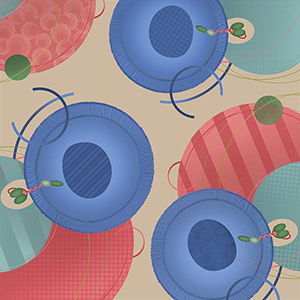
CAR T-cell product quality and stemness (Tstem) are major determinants of in vivo expansion, efficacy, and clinical response. Prolonged ex vivo culturing is known to deplete Tstem, affecting clinical outcome. YTB323, a novel autologous CD19-directed CAR T-cell therapy expressing the same validated CAR as tisagenlecleucel, is manufactured using a next-generation platform in <2 days. Here, we report the preclinical development and preliminary clinical data of YTB323 in adults with relapsed/refractory diffuse large B-cell lymphoma (r/r DLBCL; NCT03960840). In preclinical mouse models, YTB323 exhibited enhanced in vivo expansion and antitumor activity at lower doses than traditionally manufactured CAR T cells. Clinically, at doses 25-fold lower than tisagenlecleucel, YTB323 showed (i) promising overall safety [cytokine release syndrome (any grade, 35%; grade ≥3, 6%), neurotoxicity (any grade, 25%; grade ≥3, 6%)]; (ii) overall response rates of 75% and 80% for DL1 and DL2, respectively; (iii) comparable CAR T-cell expansion; and (iv) preservation of T-cell phenotype. Current data support the continued development of YTB323 for r/r DLBCL.
Significance: Traditional CAR T-cell manufacturing requires extended ex vivo cell culture, reducing naive and stem cell memory T-cell populations and diminishing antitumor activity. YTB323, which expresses the same validated CAR as tisagenlecleucel, can be manufactured in <2 days while retaining T-cell stemness and enhancing clinical activity at a 25-fold lower dose.
This study was highlighted and featured on the cover of the September issue. A related commentary is available here.
Journal: Cancer Epidemiology, Biomarkers & Prevention
Background: Individuals with adenomatous colorectal polyps undergo repeated colonoscopy surveillance to identify and remove metachronous adenomas. However, many patients with adenomas do not develop recurrent adenomas. Better methods to evaluate who benefits from increased surveillance are needed. We evaluated the use of altered EVL methylation as a potential biomarker for risk of recurrent adenomas.
Methods: Patients with ≥1 colonoscopy had EVL methylation (mEVL) measured with an ultra-accurate methylation-specific droplet digital PCR assay on normal colon mucosa. The association between EVL methylation levels and adenoma or colorectal cancer was evaluated using three case/control definitions in three models: unadjusted (model 1), adjusting for baseline characteristics (model 2), and an adjusted model excluding patients with colorectal cancer at baseline (model 3).
Results: Between 2001 and 2020, 136 patients were included; 74 healthy patients and 62 patients with a history of colorectal cancer. Older age, never smoking, and baseline colorectal cancer were associated with higher levels of mEVL (P ≤ 0.05). Each log base 10 difference in mEVL was associated with an increased risk of adenoma(s) or cancer at/after baseline for model 1 [OR, 2.64; 95% confidence interval (CI), 1.09–6.36], and adenoma(s) or cancer after baseline for models 1 (OR, 2.01; 95% CI, 1.04–3.90) and model 2 (OR, 3.17; 95% CI, 1.30–7.72).
Conclusions: Our results suggest that EVL methylation level detected in the normal colon mucosa has the potential to be a biomarker for monitoring the risk for recurrent adenomas.
Impact: These findings support the potential utility of EVL methylation for improving the accuracy for assigning risk for recurrent colorectal adenomas and cancer..
This study was highlighted in the September issue.
Journal: Cancer Immunology Research
Aerobic Exercise Alters the Melanoma Microenvironment and Modulates ERK5 S496 Phosphorylation
Exercise changes the tumor microenvironment by remodeling blood vessels and increasing infiltration by cytotoxic immune cells. The mechanisms driving these changes remain unclear. Herein, we demonstrate that exercise normalizes tumor vasculature and upregulates endothelial expression of VCAM1 in YUMMER 1.7 and B16F10 murine models of melanoma but differentially regulates tumor growth, hypoxia, and the immune response. We found that exercise suppressed tumor growth and increased CD8+ T-cell infiltration in YUMMER but not in B16F10 tumors. Single-cell RNA sequencing and flow cytometry revealed exercise modulated the number and phenotype of tumor-infiltrating CD8+ T cells and myeloid cells. Specifically, exercise caused a phenotypic shift in the tumor-associated macrophage population and increased the expression of MHC class II transcripts. We further demonstrated that ERK5 S496A knock-in mice, which are phosphorylation deficient at the S496 residue, “mimicked” the exercise effect when unexercised, yet when exercised, these mice displayed a reversal in the effect of exercise on tumor growth and macrophage polarization compared with wild-type mice. Taken together, our results reveal tumor-specific differences in the immune response to exercise and show that ERK5 signaling via the S496 residue plays a crucial role in exercise-induced tumor microenvironment changes.
A commentary related to this study is available here.
Journal: Cancer Prevention Research
Risk Factors for Early-onset Sporadic Colorectal Cancer in Male Veterans
Identifying risk factors for early-onset colorectal cancer (EOCRC) could help reverse its rising incidence through risk factor reduction and/or early screening. We sought to identify EOCRC risk factors that could be used for decisions about early screening. Using electronic databases and medical record review, we compared male veterans ages 35 to 49 years diagnosed with sporadic EOCRC (2008–2015) matched 1:4 to clinic and colonoscopy controls without colorectal cancer, excluding those with established inflammatory bowel disease, high-risk polyposis, and nonpolyposis syndromes, prior bowel resection, and high-risk family history. We ascertained sociodemographic and lifestyle factors, family and personal medical history, physical measures, vital signs, medications, and laboratory values 6 to 18 months prior to case diagnosis. In the derivation cohort (75% of the total sample), univariate and multivariate logistic regression models were used to derive a full model and a more parsimonious model. Both models were tested using a validation cohort. Among 600 cases of sporadic EOCRC [mean (SD) age 45.2 (3.5) years; 66% White], 1,200 primary care clinic controls [43.4 (4.2) years; 68% White], and 1,200 colonoscopy controls [44.7 (3.8) years; 63% White], independent risk factors included age, cohabitation and employment status, body mass index (BMI), comorbidity, colorectal cancer, or other visceral cancer in a first- or second-degree relative (FDR or SDR), alcohol use, exercise, hyperlipidemia, use of statins, NSAIDs, and multivitamins. Validation c-statistics were 0.75–0.76 for the full model and 0.74–0.75 for the parsimonious model, respectively. These independent risk factors for EOCRC may identify veterans for whom colorectal cancer screening prior to age 45 or 50 years should be considered.
Prevention Relevance: Screening 45- to 49-year-olds for colorectal cancer is relatively new with uncertain uptake thus far. Furthermore, half of EOCRC occurs in persons < 45 years old. Using risk factors may help 45- to 49-year-olds accept screening and may identify younger persons for whom earlier screening should be considered.
This study was featured on the cover of the September issue. A related commentary is available here.
Journal: Cancer Research (September 1 issue)
Genome damage is a main driver of malignant transformation, but it also induces aberrant inflammation via the cGAS/STING DNA-sensing pathway. Activation of cGAS/STING can trigger cell death and senescence, thereby potentially eliminating genome-damaged cells and preventing against malignant transformation. Here, we report that defective ribonucleotide excision repair (RER) in the hematopoietic system caused genome instability with concomitant activation of the cGAS/STING axis and compromised hematopoietic stem cell function, ultimately resulting in leukemogenesis. Additional inactivation of cGAS, STING, or type I IFN signaling, however, had no detectable effect on blood cell generation and leukemia development in RER-deficient hematopoietic cells. In wild-type mice, hematopoiesis under steady-state conditions and in response to genome damage was not affected by loss of cGAS. Together, these data challenge a role of the cGAS/STING pathway in protecting the hematopoietic system against DNA damage and leukemic transformation.
Significance: Loss of cGAS/STING signaling does not impact DNA damage–driven leukemogenesis or alter steady-state, perturbed or malignant hematopoiesis, indicating that the cGAS/STING axis is not a crucial antioncogenic mechanism in the hematopoietic system.
A commentary on this study is available here.
Journal: Cancer Research (September 15 issue)
Neoadjuvant chemoimmunotherapy (NACI) has shown promise in the treatment of resectable esophageal squamous cell carcinoma (ESCC). The microbiomes of patients can impact therapy response, and previous studies have demonstrated that intestinal microbiota influences cancer immunotherapy by activating gut immunity. Here, we investigated the effects of intratumoral microbiota on the response of patients with ESCC to NACI. Intratumoral microbiota signatures of β-diversity were disparate and predicted the treatment efficiency of NACI. The enrichment of Streptococcus positively correlated with GrzB+ and CD8+ T-cell infiltration in tumor tissues. The abundance of Streptococcus could predict prolonged disease-free survival in ESCC. Single-cell RNA sequencing demonstrated that responders displayed a higher proportion of CD8+ effector memory T cells but a lower proportion of CD4+ regulatory T cells. Mice that underwent fecal microbial transplantation or intestinal colonization with Streptococcus from responders showed enrichment of Streptococcus in tumor tissues, elevated tumor-infiltrating CD8+ T cells, and a favorable response to anti-PD-1 treatment. Collectively, this study suggests that intratumoral Streptococcus signatures could predict NACI response and sheds light on the potential clinical utility of intratumoral microbiota for cancer immunotherapy.
Significance: Analysis of intratumoral microbiota in patients with esophageal cancer identifies a microbiota signature that is associated with chemoimmunotherapy response and reveals that Streptococcus induces a favorable response by stimulating CD8+ T-cell infiltration.
A commentary on this study is available here.
Journal: Clinical Cancer Research (September 1 issue)
Purpose: Elective neck irradiation (ENI) has long been considered mandatory when treating head and neck squamous cell carcinoma (HNSCC) with definitive radiotherapy, but it is associated with significant dose to normal organs-at-risk (OAR). In this prospective phase II study, we investigated the efficacy and tolerability of eliminating ENI and strictly treating involved and suspicious lymph nodes (LN) with intensity-modulated radiotherapy.
Patients and Methods: Patients with newly diagnosed HNSCC of the oropharynx, larynx, and hypopharynx were eligible for enrollment. Each LN was characterized as involved or suspicious based on radiologic criteria and an in-house artificial intelligence (AI)–based classification model. Gross disease received 70 Gray (Gy) in 35 fractions and suspicious LNs were treated with 66.5 Gy, without ENI. The primary endpoint was solitary elective volume recurrence, with secondary endpoints including patterns-of-failure and patient-reported outcomes.
Results: Sixty-seven patients were enrolled, with 18 larynx/hypopharynx and 49 oropharynx cancer. With a median follow-up of 33.4 months, the 2-year risk of solitary elective nodal recurrence was 0%. Gastrostomy tubes were placed in 14 (21%), with median removal after 2.9 months for disease-free patients; no disease-free patient is chronically dependent. Grade I/II dermatitis was seen in 90%/10%. There was no significant decline in composite MD Anderson Dysphagia Index scores after treatment, with means of 89.1 and 92.6 at 12 and 24 months, respectively.
Conclusions: These results suggest that eliminating ENI is oncologically sound for HNSCC, with highly favorable quality-of-life outcomes. Additional prospective studies are needed to support this promising paradigm before implementation in any nontrial setting.
This article was highlighted in the September 1 issue.
Journal: Clinical Cancer Research (September 15 issue)
Purpose: To determine the role of CD49d for response to Bruton’s tyrosine kinase inhibitors (BTKi) in patients with chronic lymphocytic leukemia (CLL).
Patients and Methods: In patients treated with acalabrutinib (n = 48), CD49d expression, VLA-4 integrin activation, and tumor transcriptomes of CLL cells were assessed. Clinical responses to BTKis were investigated in acalabrutinib- (n = 48; NCT02337829) and ibrutinib-treated (n = 73; NCT01500733) patients.
Results: In patients treated with acalabrutinib, treatment-induced lymphocytosis was comparable for both subgroups but resolved more rapidly for CD49d+ cases. Acalabrutinib inhibited constitutive VLA-4 activation but was insufficient to block BCR and CXCR4–mediated inside–out activation. Transcriptomes of CD49d+ and CD49d− cases were compared using RNA sequencing at baseline and at 1 and 6 months on treatment. Gene set enrichment analysis revealed increased constitutive NF-κB and JAK–STAT signaling, enhanced survival, adhesion, and migratory capacity in CD49d+ over CD49d− CLL that was maintained during therapy. In the combined cohorts of 121 BTKi-treated patients, 48 (39.7%) progressed on treatment with BTK and/or PLCG2 mutations detected in 87% of CLL progressions. Consistent with a recent report, homogeneous and bimodal CD49d-positive cases (the latter having concurrent CD49d+ and CD49d− CLL subpopulations, irrespective of the traditional 30% cutoff value) had a shorter time to progression of 6.6 years, whereas 90% of cases homogenously CD49d− were estimated progression-free at 8 years (P = 0.0004).
Conclusions: CD49d/VLA-4 emerges as a microenvironmental factor that contributes to BTKi resistance in CLL. The prognostic value of CD49d is improved by considering bimodal CD49d expression.
A commentary on this study is available here.
Journal: Molecular Cancer Research
A major hurdle to the application of precision oncology in pancreatic cancer is the lack of molecular stratification approaches and targeted therapy for defined molecular subtypes. In this work, we sought to gain further insight and identify molecular and epigenetic signatures of the Basal-like A pancreatic ductal adenocarcinoma (PDAC) subgroup that can be applied to clinical samples for patient stratification and/or therapy monitoring. We generated and integrated global gene expression and epigenome mapping data from patient-derived xenograft models to identify subtype-specific enhancer regions that were validated in patient-derived samples. In addition, complementary nascent transcription and chromatin topology (HiChIP) analyses revealed a Basal-like A subtype-specific transcribed enhancer program in PDAC characterized by enhancer RNA (eRNA) production that is associated with more frequent chromatin interactions and subtype-specific gene activation. Importantly, we successfully confirmed the validity of eRNA detection as a possible histologic approach for PDAC patient stratification by performing RNA-ISH analyses for subtype-specific eRNAs on pathologic tissue samples. Thus, this study provides proof-of-concept that subtype-specific epigenetic changes relevant for PDAC progression can be detected at a single-cell level in complex, heterogeneous, primary tumor material.
Implications: Subtype-specific enhancer activity analysis via detection of eRNAs on a single-cell level in patient material can be used as a potential tool for treatment stratification.
This article was highlighted in the September issue.
Journal: Molecular Cancer Therapeutics

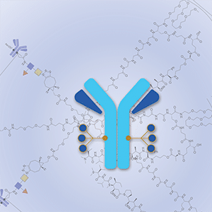
Antibody–drug conjugates (ADC) achieve targeted drug delivery to a tumor and have demonstrated clinical success in many tumor types. The activity and safety profile of an ADC depends on its construction: antibody, payload, linker, and conjugation method, as well as the number of payload drugs per antibody [drug-to-antibody ratio (DAR)]. To allow for ADC optimization for a given target antigen, we developed Dolasynthen (DS), a novel ADC platform based on the payload auristatin hydroxypropylamide, that enables precise DAR-ranging and site-specific conjugation. We used the new platform to optimize an ADC that targets B7-H4 (VTCN1), an immune-suppressive protein that is overexpressed in breast, ovarian, and endometrial cancers. XMT-1660 is a site-specific DS DAR 6 ADC that induced complete tumor regressions in xenograft models of breast and ovarian cancer as well as in a syngeneic breast cancer model that is refractory to PD-1 immune checkpoint inhibition. In a panel of 28 breast cancer PDXs, XMT-1660 demonstrated activity that correlated with B7-H4 expression. XMT-1660 has recently entered clinical development in a phase I study (NCT05377996) in patients with cancer.
This study was highlighted and featured on the cover of the September issue.
Journal: Cancer Research Communications
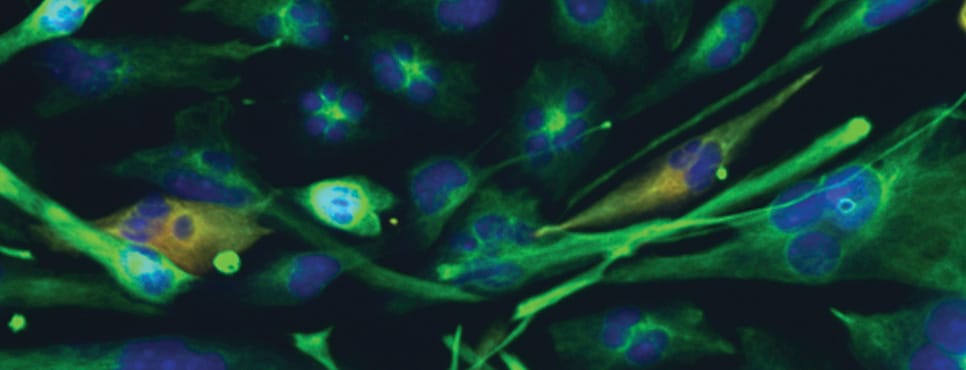
Solid cancer cells escape the primary tumor mass by transitioning from an epithelial-like state to an invasive migratory state. As they escape, metastatic cancer cells employ interchangeable modes of invasion, transitioning between fibroblast-like mesenchymal movement to amoeboid migration, where cells display a rounded morphology and navigate the extracellular matrix in a protease-independent manner. However, the gene transcripts that orchestrate the switch between epithelial, mesenchymal, and amoeboid states remain incompletely mapped, mainly due to a lack of methodologies that allow the direct comparison of the transcriptomes of spontaneously invasive cancer cells in distinct migratory states. Here, we report a novel single-cell isolation technique that provides detailed three-dimensional data on melanoma growth and invasion, and enables the isolation of live, spontaneously invasive cancer cells with distinct morphologies and invasion parameters. Via the expression of a photoconvertible fluorescent protein, compact epithelial-like cells at the periphery of a melanoma mass, elongated cells in the process of leaving the mass, and rounded amoeboid cells invading away from the mass were tagged, isolated, and subjected to single-cell RNA sequencing. A total of 462 differentially expressed genes were identified, from which two candidate proteins were selected for further pharmacologic perturbation, yielding striking effects on tumor escape and invasion, in line with the predictions from the transcriptomics data. This work describes a novel, adaptable, and readily implementable method for the analysis of the earliest phases of tumor escape and metastasis, and its application to the identification of genes underpinning the invasiveness of malignant melanoma.
Significance: This work describes a readily implementable method that allows for the isolation of individual live tumor cells of interest for downstream analyses, and provides the single-cell transcriptomes of melanoma cells at distinct invasive states, both of which open avenues for in-depth investigations into the transcriptional regulation of the earliest phases of metastasis.
Caption: This figure represents XMT-1660, a novel antibody-drug conjugate that showed potent antitumor activity in breast and ovarian cancer models.
Journal: Cancer Research Communications
Solid cancer cells escape the primary tumor mass by transitioning from an epithelial-like state to an invasive migratory state. As they escape, metastatic cancer cells employ interchangeable modes of invasion, transitioning between fibroblast-like mesenchymal movement to amoeboid migration, where cells display a rounded morphology and navigate the extracellular matrix in a protease-independent manner. However, the gene transcripts that orchestrate the switch between epithelial, mesenchymal, and amoeboid states remain incompletely mapped, mainly due to a lack of methodologies that allow the direct comparison of the transcriptomes of spontaneously invasive cancer cells in distinct migratory states. Here, we report a novel single-cell isolation technique that provides detailed three-dimensional data on melanoma growth and invasion, and enables the isolation of live, spontaneously invasive cancer cells with distinct morphologies and invasion parameters. Via the expression of a photoconvertible fluorescent protein, compact epithelial-like cells at the periphery of a melanoma mass, elongated cells in the process of leaving the mass, and rounded amoeboid cells invading away from the mass were tagged, isolated, and subjected to single-cell RNA sequencing. A total of 462 differentially expressed genes were identified, from which two candidate proteins were selected for further pharmacologic perturbation, yielding striking effects on tumor escape and invasion, in line with the predictions from the transcriptomics data. This work describes a novel, adaptable, and readily implementable method for the analysis of the earliest phases of tumor escape and metastasis, and its application to the identification of genes underpinning the invasiveness of malignant melanoma.
Significance: This work describes a readily implementable method that allows for the isolation of individual live tumor cells of interest for downstream analyses, and provides the single-cell transcriptomes of melanoma cells at distinct invasive states, both of which open avenues for in-depth investigations into the transcriptional regulation of the earliest phases of metastasis.


